HoloWrist
An FWO Research Project
The Challenge
Improving Wrist Surgery
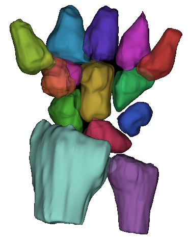
When you break the scaphoid bone in your wrist, it is common to surgically install a screw to bring the broken pieces of bone back together. The video on the right shows this surgery taking place.
It is crucial to place the screw as accurately as possible. Failure to position the screw accurately can lead to longer surgeries and poor bone healing. This goal is complicated by the factors listed below.
Complicating Factors
To improve wrist surgery outcomes, three current limiting factors need to be addressed.
Obstructed Views
Because of the wrist's complex anatomy, and the precarious blood supply to
bones in the wrist, any incision for surgical fixation of a fracture has
to be small. This small incision requirement
makes it so that the broken wrist bone cannot be directly observed by the
surgeon.
Radiation
Because the surgeon cannot see the broken bone with their own eyes, x-ray
imaging (fluoroscopy) is used to see the bones during the surgery. Unfortunately,
x-ray imaging produces harmful radiation, which means that it can only be used
sparingly.
2D to 3D Mapping
While x-ray gives the surgeon information on the position of the broken wrist bone, that information is in 2D and shown on a separate monitor. The surgeon then has the complicated task of interpreting those 2D x-ray images and transferring that information to the real 3D wrist of the patient.

Follow Our Progress
You can follow our progress through the social media links below. This research project is a collaboration between Ghent University, KU Leuven, and the MoRe Institute. Funding is provided by the Flanders Research Foundation (FWO).
|
|
|
|
|